Panoramic X-rays and the art of planning dental implants
Every dental implant procedure begins with an image. Before a drill touches bone, the surgeon needs to know what lies beneath the gum: how thick the ridge is, where the nerve curves through the lower jaw, and how far the sinus dips into the upper arch. The panoramic X-ray — also called an orthopantomogram, or OPG — has been the workhorse of this early assessment for decades, and it still does the job better than most people realise.
But a flat image is only ever a sketch, not a blueprint. Modern implantology is digital, three-dimensional and driven by the final tooth rather than the empty socket, which raises a fair question about where two-dimensional imaging still fits. The honest answer is that a panoramic radiograph earns its place at the start of the process: it surveys the whole mouth, flags what nobody was expecting, and provides the baseline against which everything else is measured. Knowing its role and its limits helps patients turn up to a consultation ready to ask sensible questions.
What a panoramic X-ray shows you (and what it doesn't)
An OPG sweeps an X-ray source and detector around the head in a single pass, capturing both jaws, the sinuses and the neck in one flattened frame. In Australian practices it is quick, comfortable and comparatively cheap. Most clinics take one within minutes of a new patient examination, and it remains the standard first-line survey before anything invasive is discussed.
What it reveals is genuinely useful. The entire arch and both sinus floors appear at once, so a surgeon can spot a retained root, a cyst, an impacted wisdom tooth, a fractured existing fixture or bone resorption that changes the plan entirely. It is also the best single view for judging gross ridge volume and comparing the left side with the right.
The limitation is geometric. A panoramic image is a projection, so magnification varies across the frame: teeth and bone outside the narrow focal trough can look stretched or squeezed, and structures in the anterior region blur together. It is a poor measuring instrument. Estimating bone height above the inferior alveolar nerve or the depth of a sinus from a panoramic film carries an error margin large enough to matter. Experienced clinicians straighten the arch in the software to reduce that error, but they never treat the numbers as gospel.
Why implant planning starts wider than a single tooth
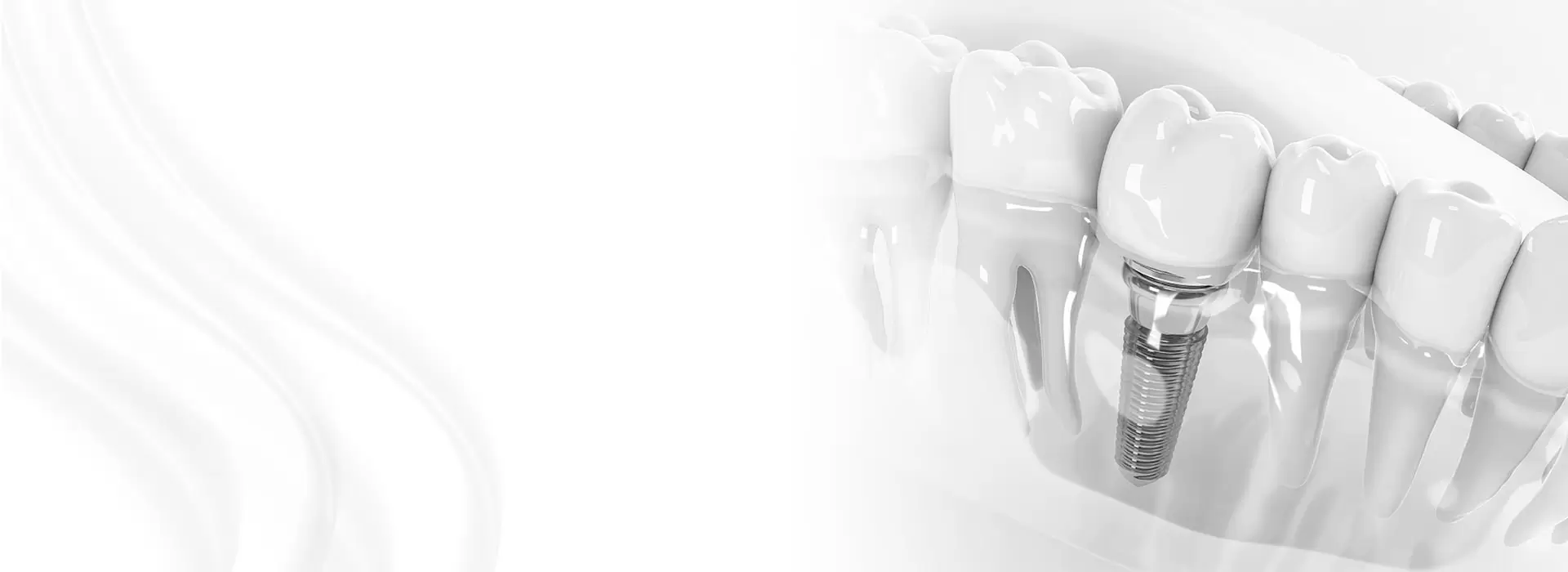
Implant placement is close to irreversible. The position of the fixture in bone dictates where the crown sits, and the crown is what the patient actually judges the result on. This is the prosthetically driven principle: design the finished tooth first, then work backwards to the bone. Doing that requires the height and width of the ridge, the location of the mental foramen and the nasopalatine canal, the shape of any sinus septa, the thickness of the cortical plates and, increasingly, the density of the bone that will hold the implant.
This is where the panoramic image shifts from decision tool to screening tool. It tells the team the case is complex; a cone-beam CT then supplies the numbers. Australian practice is broadly conservative on this point — CBCT is justified when two-dimensional imaging cannot answer the question, not simply because the machine sits in the corner of the room. The dose gap is real. A limited-field CBCT of one quadrant delivers a fraction of the radiation of a full medical CT but several times that of a panoramic scan, so it is ordered deliberately.
There is a local wrinkle worth knowing. Medicare does not subsidise implant surgery, so patients fund it themselves or through private cover with a major dental policy, and money is usually the first thing discussed. Geography matters too. Someone driving in from Bendigo for a full-arch rehabilitation, or travelling from the Hunter Valley down to Sydney, cannot keep coming back each time a new image is needed. One well-planned panoramic study, combined with a surgical guide, keeps those trips down to a single visit.
From scan to surgical guide
The workflow that follows imaging is where planning becomes real. Digital intraoral scanners capture teeth and gums in three dimensions; those files are merged with the CBCT volume in planning software, where the technician positions the implant, checks safety margins of a couple of millimetres from the nerve and the sinus, and generates a stereolithographic surgical guide the surgeon drills through. The result is millimetre precision instead of a freehand guess.
Panoramic imaging still appears in that workflow, usually earlier and for a broader purpose. It confirms the jaws are free of pathology before surgery starts, and it gives the surgeon a mental map of the whole arch rather than one quadrant, which is exactly what a full-mouth rehabilitation demands.
Clinics that work this way build the plan around the finished result and the person in front of them. In Izmir, the team at TruDent Clinics pairs panoramic assessment with digital planning and individualised treatment plans across implants, grafting and same-day restorative work, an approach described on TruDent's clinical site. That sequence is worth discussing before anyone travels abroad for treatment, because the number of visits required depends heavily on how thoroughly imaging is handled in the first sitting.
Getting ready for your implant assessment
Imaging is technical, but preparation is mostly administrative. Patients who arrive with organised records tend to leave with a sharper plan and fewer repeat appointments.
- Bring any previous X-rays, CT volumes or surgical guides from other practices; they are often more useful than repeating identical imaging.
- Ask for the panoramic image as a digital file rather than a printed film, so it can be merged into planning software and re-measured.
- Request a surgical guide wherever the anatomy allows; it shortens surgery and reduces the chance of a misplaced implant.
- Get a written plan naming the implant system, the number of fixtures, the grafting and the timeline, so quotes from different clinics can be compared honestly.
- Clarify who handles the graft, the implant and the crown, because in Australia these are sometimes split between a specialist surgeon and a restorative dentist.
- Ask whether a limited-field CBCT is genuinely needed and who will order it, so the cost and the radiation exposure are clear before you consent.
Imaging options compared side by side
Not every patient needs every scan. The choice depends on how much the team already knows and how complex the case looks after a clinical examination, so the following comparison is worth keeping in mind when a clinic proposes a pathway.
| Imaging method |
What it reveals |
Role in implant planning |
Notes for Australian patients |
| Periapical radiograph |
One tooth, its root and surrounding bone in fine detail |
Baseline for a single site; bone level around neighbouring teeth |
Small field, low dose, often bulk-billed |
| Bitewing radiograph |
Crowns of the upper and lower back teeth and the bone crest |
Checks the bone that will support crowns and existing fixtures |
Quick, low dose, routine at recall visits |
| Panoramic radiograph (OPG) |
Both jaws, sinuses, full arch and nerve canal outline |
First-line survey; uncovers pathology that alters the plan |
Widely available, same-day, reasonably priced |
| Limited-field CBCT |
True 3D bone, nerve canal and sinus anatomy |
Precise measurement, implant positioning, guide fabrication |
Higher dose than a panoramic scan; usually an extra fee |
| Full medical CT |
Entire head and soft tissue |
Rarely justified for routine implant work |
Considerably higher dose, reserved for complex cases |
The panoramic study usually comes first because it is fast and answers the screening question outright. The CBCT follows only when exact measurements are actually needed, which keeps radiation and out-of-pocket costs down for straightforward cases.
Book a comprehensive implant consultation
The most valuable first appointment is a proper assessment, not a sales pitch. Ask what imaging will be taken, who interprets it, whether a surgical guide is planned, and what the quoted total covers — grafting, the crown, and any revision visit. Patients in Melbourne, Sydney and Brisbane can usually complete the panoramic scan and clinical examination in a single sitting, and clinics will supply the scan digitally for your records. If you are weighing treatment at home against travelling to see a specialist, the same checklist applies: request the imaging and the written plan before booking flights, so the trip is worth the money.
Send your previous scans and a current list of medications with the booking request, since blood thinners and certain supplements affect both imaging and surgery. TruDent Clinics offers consultation-led implant planning alongside cosmetic and general dentistry, so a full-mouth assessment can be arranged in one place rather than pieced together across separate providers.