Why a 3D Imaging Consultation Sets the Tone for Better Dentistry
For anyone weighing up veneers, implants, or a full smile makeover, the very first appointment shapes the entire experience. A consultation built around 3D imaging goes beyond the standard visual check and a flat bitewing X-ray, giving patients and clinicians a precise digital model of the teeth, jaw, and surrounding structures from the moment they sit in the chair.
Australian patients often begin their research from a dentist in Sydney or Melbourne, only to realise that local quotes for complex cosmetic work can stretch well into five figures. That reality pushes many to look overseas, where modern clinics in Turkey now offer diagnostic consultations powered by cone-beam CT scans and intraoral scanners. Understanding what those advanced appointments deliver helps cut through the noise before booking flights from Tullamarine or Kingsford Smith.
What a 3D Imaging Consultation Actually Involves
A 3D imaging consultation starts with a cone-beam computed tomography scan, which rotates around the head and captures hundreds of layered images in a single sweep. These are stitched together into a three-dimensional model that can be rotated, sliced, and measured on a screen in real time. Most appointments also include a digital intraoral scan, replacing the messy impression trays that patients remember from past visits.
The dentist walks through the model with the patient, pointing out bone density, root positions, sinus cavities, and the way the upper and lower jaws meet. For anyone who has ever had a dentist say "open a little wider" while squinting at a two-dimensional film, the upgrade feels significant. The scan is usually finished within a minute, and the software does the heavy lifting in the background.
Traditional vs 3D Imaging: A Side-by-Side Look
The differences between a standard consult and a 3D imaging consult show up quickly once you line them up.
| Feature |
Standard consultation |
3D imaging consultation |
| Imaging method |
2D bitewing or panoramic X-ray |
Cone-beam CT plus intraoral scan |
| Bone and root detail |
Flat, overlapping view |
Three-dimensional, rotatable model |
| Impressions |
Putty or alginate trays |
Digital intraoral scan |
| Treatment simulation |
Verbal description or mock-up |
Software-based smile preview |
| Suitability for implants |
Limited |
Full pre-planning with guided surgery |
| Time on site |
Often a single short visit |
Scan plus planning session |
| Records shared remotely |
Low-resolution photos |
Full digital file set |
The extra data translates into fewer assumptions on treatment day. Patients in regional Queensland or Western Australia who may only be able to travel once often find this especially useful, as every minute of the trip can be used for confirmed treatment rather than re-diagnosis.
Catching Aesthetic and Functional Issues Before They Worsen
A 3D scan picks up problems that flat X-rays routinely miss. Hairline fractures in roots, early bone loss around molars, the exact position of an impacted wisdom tooth, and the relationship between upper teeth and sinus floor are visible without invasive exploration. This matters most when planning treatments that affect appearance as well as function, and resources that explain aesthetic and functional deficiencies outline exactly why these subtle issues often dictate the final treatment plan.
Because the scan captures the bite from multiple angles, clinicians can also spot uneven wear, TMJ strain, and small airway restrictions that contribute to grinding or snoring. Addressing these alongside cosmetic work avoids the all-too-common outcome where the smile looks great in photos but the patient still suffers headaches or jaw fatigue months later.
Precision Planning for Implants, Veneers, and Crowns
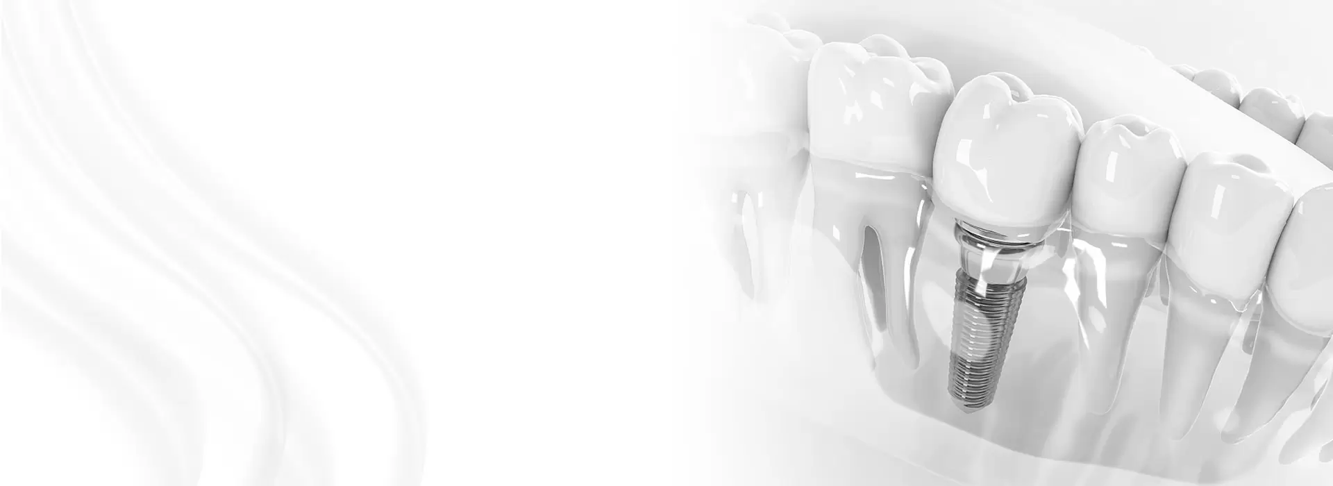
For dental implants, a 3D consultation is now considered best practice. The scan shows bone width, height, and density, allowing the surgeon to place the implant virtually before picking up a drill. A surgical guide can be 3D-printed from that plan, so the actual placement mirrors the digital rehearsal within a fraction of a millimetre. Veneer and crown planning benefit too, as the digital model feeds straight into CAD software that designs the final restorations.
This level of precision changes the conversation between dentist and patient. Instead of a rough estimate of how many veneers might work, the patient sees a rendered preview on their own digital smile. Adjustments to shape, length, and shade happen on screen before anything is milled, which removes much of the guesswork from cosmetic work.
Reducing Repeat Visits and Unexpected Surprises
One of the hidden costs of any dental treatment is the repeat visit. A crown that does not seat properly, an implant placed too close to a nerve, a veneer shade that looks wrong in daylight — all of these force patients back into the chair. With 3D imaging, the most common surprises are caught during planning rather than on the day of treatment.
For Australians travelling overseas for dental work, fewer surprises matter even more. Most people can manage a single trip of seven to ten days, but coordinating follow-up visits from the other side of the world is awkward and expensive. A thorough 3D consultation beforehand trims the margin for error and keeps the trip on schedule, even when the patient boards a plane home feeling relieved rather than anxious.
Confidence for Australian Patients Considering Treatment Abroad
Trust is the biggest hurdle for Australians looking at overseas dental care, and rightly so. The Therapeutic Goods Administration regulates what is used in Australia, and stories about botched overseas work have put many people off the idea entirely. A consultation that includes 3D imaging gives patients something tangible to assess before committing. They can review the digital scan, see the proposed outcome rendered in software, and ask informed questions about materials, implant brands, and timelines.
This transparency also makes it easier to compare clinics fairly. Some Australian patients cross-check their scans with a local dentist before flying, while others send their files to a second clinic for an opinion. Reading about the experience at Dr Cetin Clinic gives a clear picture of how a serious Turkish practice presents its diagnostic process, and that kind of reference point helps separate marketing from substance.
Long-Term Value Through Digital Records
The files produced during a 3D imaging consultation do not disappear when the appointment ends. The CBCT scan, intraoral model, photos, and treatment plan become part of a permanent digital record. If a veneer chips five years later, the original model is still on file, and a replacement can be milled to match. If an implant crown needs adjustment, the surrounding structures are already mapped.
This kind of continuity is increasingly rare in dentistry. Many Australian patients find that records from a previous practice are difficult to retrieve, especially after a change of clinic. A digital-first workflow sidesteps the paperwork entirely. The result is a treatment journey that begins with clarity and continues with consistency, which is exactly what most patients want when investing in their smile.
If a consultation with 3D imaging sounds like the right starting point, the next step is a simple online enquiry. TruDent Clinics offers personalised treatment planning built around advanced digital diagnostics, and the team can arrange a remote review of any existing scans before flights are even booked. Reach out today and find out whether the trip from Sydney, Perth, or anywhere in between could end in a smile worth showing off at every footy match from now on.